
18
Jul
Radiology and Scanning: Why Imaging Matters in Early Diagnosis
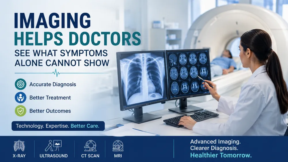
When someone experiences pain, swelling, fever, breathlessness, or an injury, the first step in diagnosis is usually a detailed medical history and physical examination. While these provide valuable information, they do not always reveal what is happening inside the body. Many medical conditions cannot be accurately diagnosed based on symptoms alone. This is where radiology and diagnostic imaging become essential.
Modern imaging technologies allow doctors to look inside the body safely and non-invasively, helping them identify the exact cause of a patient's symptoms. Whether it is a fractured bone, kidney stone, pregnancy assessment, lung infection, or abdominal condition, imaging often provides the clarity needed for an accurate diagnosis and appropriate treatment.
Many patients feel anxious when their doctor recommends an X-ray or ultrasound scan. Some worry that the problem may be serious, while others wonder if the scan is truly necessary. In reality, imaging is usually requested not because doctors expect the worst, but because they want to make informed decisions based on clear evidence rather than assumptions.
For people living in Mattannur, Kannur, Kuthuparamba, Irikkur, Kasaragod, and nearby areas, access to reliable radiology services helps doctors diagnose medical conditions quickly, monitor treatment progress, and provide better patient care.
What Is Radiology?
Radiology is the branch of medicine that uses medical imaging to diagnose, monitor, and sometimes even guide the treatment of diseases and injuries.
Instead of relying solely on external symptoms, radiology allows healthcare professionals to visualize organs, bones, blood vessels, muscles, and other internal structures.
Different imaging methods are used depending on the medical condition being investigated. Each technique has its own purpose, advantages, and limitations.
The goal is always to obtain the most useful information while minimizing unnecessary testing.
Why Diagnostic Imaging Is Important
Many health conditions produce similar symptoms.
For example, abdominal pain could result from:
- Kidney stones
- Gallbladder disease
- Appendicitis
- Liver disorders
- Intestinal problems
- Urinary tract conditions
- Ovarian disorders
- Muscle strain
Without imaging, it may be difficult to determine the exact cause.
Similarly, chest pain may originate from the lungs, heart, muscles, ribs, or digestive system. Imaging helps narrow down the possibilities and supports accurate diagnosis.
Rather than replacing clinical examination, imaging complements it by providing objective information that cannot be obtained through physical examination alone.
Common Types of Diagnostic Imaging
Several imaging techniques are commonly used in everyday medical practice.
X-ray
X-rays are among the oldest and most widely used imaging methods.
They are particularly useful for examining bones and certain chest conditions.
Doctors commonly request X-rays to evaluate:
- Bone fractures
- Joint injuries
- Arthritis
- Chest infections
- Lung conditions
- Spine problems
- Dental issues
X-rays are quick, painless, and often completed within a few minutes.
Ultrasound
Ultrasound uses sound waves rather than radiation to produce images of internal organs.
It is considered extremely safe and is widely used for many medical conditions.
Ultrasound may be recommended for:
- Pregnancy monitoring
- Liver disease
- Gallbladder stones
- Kidney stones
- Urinary bladder problems
- Thyroid disorders
- Pelvic conditions
- Soft tissue swelling
- Blood vessel evaluation
Because ultrasound does not use ionizing radiation, it is especially valuable during pregnancy.
CT Scan
Computed Tomography (CT) provides highly detailed cross-sectional images of the body.
CT scans are often recommended when doctors require more detailed information than an ordinary X-ray can provide.
They may help evaluate:
- Head injuries
- Brain disorders
- Internal bleeding
- Lung disease
- Complex fractures
- Abdominal emergencies
- Certain cancers
CT scans are particularly useful during emergency situations because they provide rapid and comprehensive imaging.
MRI
Magnetic Resonance Imaging (MRI) uses magnetic fields and radio waves to create highly detailed images.
MRI is particularly valuable for examining:
- Brain
- Spine
- Ligaments
- Muscles
- Joints
- Soft tissues
- Certain neurological conditions
MRI does not use ionizing radiation, although the procedure usually takes longer than an X-ray or CT scan.
When Does Your Doctor Recommend Imaging?
Doctors recommend imaging only when it is likely to improve diagnosis or treatment planning.
Some common situations include:
Bone and Joint Injuries
Falls, sports injuries, road accidents, or sudden swelling may require imaging to identify fractures, ligament injuries, or joint damage.
An X-ray often confirms broken bones, while MRI may be necessary for ligament or cartilage injuries.
Persistent Abdominal Pain
Pain in the abdomen can have many causes.
Ultrasound often helps evaluate:
- Gallstones
- Kidney stones
- Liver disease
- Pancreatic disorders
- Pelvic conditions
- Urinary tract problems
Early diagnosis helps avoid unnecessary complications.
Pregnancy Care
Ultrasound plays an important role throughout pregnancy.
It helps monitor:
- Baby's growth
- Heartbeat
- Placental position
- Amniotic fluid
- Fetal development
- Pregnancy complications
Routine pregnancy scans provide valuable information that supports safe maternal and fetal care.
Chest Symptoms
Persistent cough, chest pain, fever, breathlessness, or suspected pneumonia often require chest imaging.
Chest X-rays help doctors evaluate infections, lung diseases, and certain heart conditions.
Kidney Stones
Severe flank pain, painful urination, or blood in the urine may indicate kidney stones.
Ultrasound or CT imaging helps identify stone location, size, and associated complications.
Liver and Gallbladder Conditions
Patients experiencing abdominal discomfort, jaundice, nausea, or abnormal liver function tests may benefit from ultrasound evaluation.
Imaging helps identify fatty liver, gallstones, liver enlargement, cysts, and other abnormalities.
Imaging Supports Better Treatment Decisions
One of the greatest benefits of diagnostic imaging is improved treatment planning.
Rather than relying on assumptions, doctors receive visual confirmation of the underlying condition.
This allows them to:
- Confirm diagnosis
- Rule out serious conditions
- Select appropriate treatment
- Decide whether surgery is necessary
- Monitor healing
- Evaluate treatment response
Accurate diagnosis often prevents unnecessary medications and procedures.
Is Medical Imaging Safe?
Many patients worry about the safety of imaging tests.
The answer depends on the type of scan being performed.
X-rays and CT Scans
These imaging techniques use carefully controlled doses of radiation.
The radiation exposure from medically indicated imaging is generally considered safe when used appropriately.
Doctors recommend these tests only when the expected diagnostic benefit outweighs any potential risk.
Ultrasound
Ultrasound uses sound waves rather than radiation.
It is widely regarded as one of the safest imaging techniques and is routinely used during pregnancy.
MRI
MRI uses magnetic fields instead of radiation.
Although the examination takes longer, it is considered safe for most patients, except those with certain implanted medical devices or metal objects.
Why Unnecessary Imaging Should Be Avoided
While diagnostic imaging is extremely valuable, not every symptom requires a scan.
Ordering unnecessary imaging may:
- Increase healthcare costs
- Delay appropriate treatment
- Lead to incidental findings that create unnecessary anxiety
- Expose patients to avoidable radiation in some cases
This is why doctors carefully evaluate each patient's symptoms before recommending any imaging study.
The most appropriate scan depends on the individual's medical condition, age, examination findings, and clinical history.
Preparing for Your Imaging Appointment
Preparation varies depending on the type of imaging.
Patients may be advised to:
- Wear comfortable clothing
- Remove jewelry or metal objects
- Avoid eating before certain scans
- Drink water before some ultrasound examinations
- Inform staff about pregnancy
- Carry previous imaging reports if available
Following preparation instructions helps improve image quality and diagnostic accuracy.
The Role of Experienced Radiologists
Medical images must be interpreted accurately.
Radiologists are specially trained doctors who analyze imaging studies and identify abnormalities that may not be obvious to others.
Their detailed reports help treating physicians combine imaging findings with clinical examination and laboratory results to reach the correct diagnosis.
Close collaboration between radiologists and other specialists leads to more effective patient care.
Modern Radiology Improves Patient Outcomes
Advances in medical imaging have transformed healthcare.
Today, many conditions that once required exploratory surgery can now be diagnosed quickly through imaging alone.
Radiology helps reduce unnecessary procedures, shortens diagnosis time, improves treatment planning, and supports better recovery.
From emergency medicine to pregnancy care, orthopedics, internal medicine, surgery, and preventive healthcare, imaging plays a central role in nearly every medical specialty.
Radiology Services at HNC Hospitals
At HNC Hospitals, radiology services support accurate diagnosis through modern imaging technology and experienced medical professionals. Diagnostic imaging is used to evaluate a wide range of conditions, including bone injuries, abdominal pain, pregnancy monitoring, liver and gallbladder disorders, kidney stones, chest conditions, and other medical concerns.
Every imaging study is recommended based on clinical need, ensuring that patients receive the most appropriate investigation for their symptoms. The radiology team works closely with physicians and specialists to provide timely reports that assist in accurate diagnosis and effective treatment planning.
By combining advanced imaging facilities with patient-centered care, HNC Hospitals helps patients receive reliable diagnostic support while minimizing unnecessary investigations.
Conclusion
Many illnesses and injuries cannot be fully understood through symptoms alone. Diagnostic imaging allows doctors to safely examine the body's internal structures, making it easier to identify the exact cause of a patient's condition. Whether evaluating fractures, pregnancy, abdominal pain, kidney stones, lung disease, or soft tissue injuries, radiology plays a vital role in modern healthcare.
Medical imaging is not meant to replace clinical examination—it enhances it by providing objective information that leads to more accurate diagnoses and better treatment decisions. When used appropriately and interpreted by experienced professionals, imaging improves patient outcomes, reduces uncertainty, and helps ensure timely medical care.
If your doctor recommends an imaging study, it is usually an important step toward understanding your condition more clearly and receiving the right treatment at the right time.

